What happens when microbial biotechnology meets material engineering? A PROMICON-funded research
A PROMICON-supported research article has been recently published in the Society for Applied Microbiology (sfam) Journal. The study investigates the outcomes of the interaction between microbial biotechnology and material engineering and has been conducted by a team of leading experts at the Spanish National Research Council (CSIC).
Various branches of bacterial biopolymers have been attracting the interest of researchers in many fields, for instance, biomedicine and packaging, due to their biodegradable, biocompatible and renewable nature. The emergence of new technologies, such as synthetic biology, enables the creation of next-generation advanced materials presenting smart functional properties, for example, the ability to sense and respond to stimuli as well as the capacity for self-repair. Herein, the research team has investigated how the newly engineered living materials (ELMs) can be ‘brought to life’ through the incorporation of living organisms.
The study has highlighted the enormous non-native diversity that can be generated from bacterial biopolymers through the synergistic combination of microbial biotechnology, synthetic biology, metabolic engineering, and materials science. The results of the research show that current trends point to the potential for bacterial biopolymers for use as scaffolds, which harbour living organisms, with the ultimate goal of creating ELMs, in which the material can self-repair, detect and respond to stimulation.
The full paper you can read here.
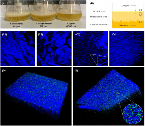
Photo: A. Bacterial cellulose being produced in static culture by three strains of the genus Komagataeibacter that yield different amounts of the biopolymer. B. 3D structure of BC in static culture. C-1 to C-4. Confocal laser scanning microscopy images acquired at the different depths of the BC membrane set in image B. D and E. Different views of a 3D reconstructed structure of a BC membrane being created by K. medellinensis. Images C–E were acquired through confocal laser scanning microscopy using Calcofluor White M2R to stain cellulose, which was coloured in blue and LIVE/DEAD™ BacLight™ kit having SYTO 9 and propidium iodide to stain living and dead cells, coloured in green and red, respectively.